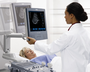
 Ultrasound imaging, also called ultrasound scanning or sonography, involves exposing part of the body to high-frequency sound waves to produce pictures of the inside of the body. Ultrasound exams do not use ionizing radiation (x-ray). Because ultrasound images are captured in real-time, they can show the structure and movement of the body’s internal organs, as well as blood flowing through blood vessels.
Ultrasound imaging is usually a painless medical test that helps physicians diagnose and treat medical conditions.
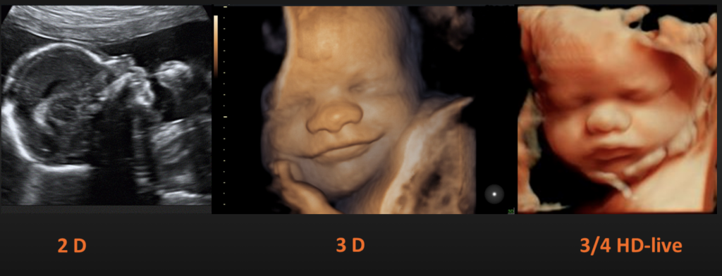
“4D” is shorthand for “four-dimensional”- the fourth dimension being time. As far as ultrasound is concerned, 4D Ultrasound is the latest ultrasound technology. 4D Ultrasound takes three-dimensional still ultrasound images and adds the element of time to the process. The result: live action images of your unborn child.
As with all ultrasound systems, it can be used to research the following
- Determining the age of the baby
- Analyzing development of the baby
- Evaluating multiple pregnancies
- Detecting structural problem with uterus
- Detecting placental abnormalities
- Detecting abnormal bleeding
- Determining ectopic pregnancy
- Detecting ovarian tumor/fibroids
- Locating the placenta
 Elastography uses low frequency vibrations during an ultrasound or MRI to measure the stiffness (or elasticity) of organs inside the body. It is particularly useful for detecting the presence and severity of liver disease.
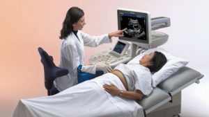
 Most ultrasound examinations are painless, fast and easy.After you are positioned on the examination table, the radiologist or sonographer will spread some warm gel on your skin and then press the transducer firmly against your body, moving it back and forth over the area of interest until the desired images are captured. There may be varying degrees of discomfort from pressure as the transducer is pressed against the area being examined. If scanning is performed over an area of tenderness, you may feel pressure or minor pain from the procedure.
At times the sonographer may have to press more firmly to get closer to the embryo or fetus to better visualize the structure. Any discomfort is usually minimal and temporary. If a Doppler ultrasound study is performed, you may actually hear pulse-like sounds that change in pitch as the blood flow is monitored and measured. With transvaginal scanning, there may be minimal discomfort as the transducer is moved in the vagina, especially when the bladder begins to refill. Once the imaging is complete, the gel will be wiped off your skin.After an ultrasound exam, you should be able to resume your normal activities.
 Lakes Radiology is an Accredited Facility with the American College of Radiology. ACR Accreditation is recognized as the gold standard in medical imaging since 1987.
The American College of Radiology® (ACR®) is a nonprofit professional society representing radiologists, nuclear medicine physicians, radiation oncologists and medical physicists. It is the largest and oldest imaging accrediting body in the U.S., with a current membership of 39,000 physicians and medical physicists. The core purpose of the ACR is to serve patients and society by empowering its members to advance the practice, science and professions of radiological and radiation oncology care.