 CT (computed tomography), sometimes called CAT scan, uses special x-ray equipment to obtain image data from different angles around the body and then uses computer processing of the information to show a cross-section of body tissues and organs.
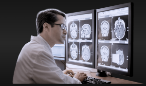
CT imaging is particularly useful because it can show several types of tissue — lung, bone, soft tissue and blood vessels—with great clarity. Using specialized equipment and expertise to create and interpret CT scans of the body, radiologists can more easily diagnose problems such as cancers, cardiovascular disease, infectious disease, trauma and musculoskeletal disorders.
Lakes Radiology has the latest technology in (MDCT) CT scanning, this revolutionary system is emerging as a highly effective screening tool in the early detection of heart disease and other serious medical conditions — even years before major symptoms occur.
Our GE LightSpeed Plus High Speed Multislice CT Scanner allows very fast scanning.. so fast in fact that most scans can be performed in less than a minute. these subsecond scans result in much improved quality because of decresased patient motion and fast adquisitions, as usual we have choosen a patient friendly scanning of all patients, large, small and claustrophobic or not.
 The technologist begins by positioning the patient on the CT table. The patient’s body may be supported by pillows to help hold it still and in the proper position during the scan. As the study proceeds, the table will move slowly into the CT scanner. Depending on the area of the body being examined, the increments of movement may be so small that they are almost undetectable or large enough that the patient feels the sensation of motion.
A CT examination often requires the use of different contrast materials to enhance the visibility of certain tissues or blood vessels. The contrast material may be swallowed, injected through an IV directly into the blood stream or administered by enema, depending on the type of examination. Before administering the contrast material, the radiologist or technologist may ask whether the patient has any allergies, especially to medications or iodine, and whether the patient has a history of diabetes, asthma, a heart condition, kidney problems or thyroid conditions. These conditions may indicate a higher risk of reaction to the contrast material or potential problems eliminating the material from the patient’s system after the exam.
A CT examination usually takes five minutes to half an hour. When the exam is over the patient may be asked to wait until the images are examined to determine if more images are needed.
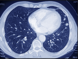
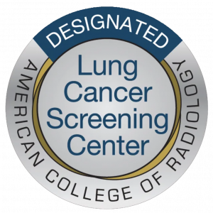 Lakes Radiology is an accredited facility with the American College of Radiology to perform the low dose lung CT, the only recommended screening test for lung cancer. This test is a low-dose computed tomography exam (also called a low-dose CT scan, or LDCT).
The American College of Radiology® (ACR®) is a nonprofit professional society representing radiologists, nuclear medicine physicians, radiation oncologists and medical physicists. It is the largest and oldest imaging accrediting body in the U.S., with a current membership of 39,000 physicians and medical physicists. The core purpose of the ACR is to serve patients and society by empowering its members to advance the practice, science and professions of radiological and radiation oncology care.