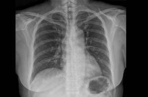
 X-rays (also called radiographs) are produced by passing a small amount of controlled radiation through the body. Radiation from x-rays are absorbed differently by the tissues of the body. When the x-rays pass through the body, these differences are captured on a special film plate that is placed behind the patient. For example, bone absorbs more radiation than soft tissue, making it appear bright white on film. Radiologists use x-ray films to detect and help diagnose certain conditions such as broken bones, pneumonia, and emphysema.
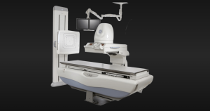
Lakes Radiology offers a wide range of x-ray procedures . We have three systems under the same roof, giving us the ability to accommodate a large amount of patients and keep the wait time minimal.
 This technology is currently very new. These plates trap the x-ray energy and require an intermediate processing step to release the stored information so it can be converted into a digital picture. Just as many recordings and music albums now sound sharper and better than ever on a digital compact disk (CD) player than on and older (analog) record player, digital x-rays look sharper and cleaner than the analog version.
Benefits of digital technology to all x-ray systems include:
- Lower dosage x-rays can often be used to achieve the same high quality picture as with film
- Digital x-ray images can be enhanced and manipulated with computers and sent via a network to other workstations and computer monitors so that many people can share the information and assist in the diagnosis.
- Digital images can be archived onto compact optical disks or digital tape drives saving tremendously on storage space and manpower needed for a traditional x-ray film library
- Digital images may be retrieved from an electronic archive for future reference
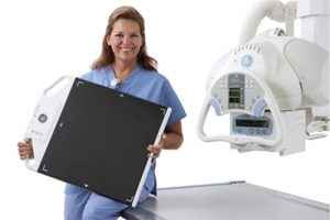 Before your x-ray, you may be asked to change into an examination gown and to remove any metal from your body (eg, eyeglasses, jewelry, watch). Next, you will be taken into the x-ray suite and positioned on an examination table. Once you are in position, the technologist will leave the room and may ask you to hold your breath if x-rays are taken of the chest. Holding your breath is very important because motion of the lungs during regular breathing can blur the images. Next, the technologist will come back into the room to put new film in the machine and you will most likely be asked to change position to allow imaging from different viewpoints.
After the x-rays have been taken, they will be developed and briefly reviewed by the radiologist to make sure that more images are not needed. You will then be asked to change back into your clothes.
After the radiologist has studied your x-ray films in more detail, a report will be sent to your referring physician, who will discuss these results with you and determine a course of action.
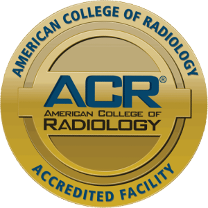 Lakes Radiology is an Accredited Facility with the American College of Radiology. ACR Accreditation is recognized as the gold standard in medical imaging since 1987.
The American College of Radiology® (ACR®) is a nonprofit professional society representing radiologists, nuclear medicine physicians, radiation oncologists and medical physicists. It is the largest and oldest imaging accrediting body in the U.S., with a current membership of 39,000 physicians and medical physicists. The core purpose of the ACR is to serve patients and society by empowering its members to advance the practice, science and professions of radiological and radiation oncology care.