 Digital Breast Tomosynthesis (DBT) acquires a series of low-dose images at different angles, the acquired images are reconstructed into a series of high-resolution slices displayed individually or dynamically in a cine mode making the Hexagonal pixels distribute the electrical field more efficiently than traditional square pixels, to capture stronger signals with less noise obtaining exceptionally sharp images, even at low dose.
3D Digital Mammography with Tomosynthesis (DBT) offers shorter examination times and significantly improved patient comfort and convenience since the time the patient must remain still is much shorter.
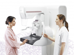
At Lakes Radiology we offer 3D Digital mammography that produces high-resolution images with low X-ray dose, Dual mode Tomosynthesis and comfort functions. Tomosynthesis makes it possible to observe the internal structure of the breast.
The images taken from different angles are reconstructed into a range of Tomosynthesis slices where the structure of interest is always in focus.
The reconstructed tomographic images make it easier to identify lesions which might be difficult to visualize in routine mammography because of the presence of overlapping breast structures.
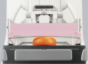 This function will reduce the compression pressure within a range (within + 3 mm) in which the thickness of the breast does not change after normal breast compression is completed for the purpose of alleviating the patient’s pain. For breast compression, there is a phenomenon (hysteresis*1) where the thickness of the breast becomes thinner during decompression after compression than during compression even with the same pressure. By utilizing this phenomenon, it is possible to automatically decompress it so that the breast condition remains almost the same even if the duration of maximum compression pressure is reduced.
 Patient preparation involves removing any articles of clothing or jewelry that might interfere with the creation of the x-ray image (see above ” Preparations for Mammogram” list for more information).
- During mammography, a technologist will position the patient and image the breast. The breast is first placed on a special cassette and gently compressed with a paddle (often made of clear Plexiglas or other plastic). This flattens the breast so that the maximum amount of tissue can be imaged and examined. Breast compression may cause some discomfort, but only lasts for a brief time during the mammography procedure .
- The technologist will step behind a special shielded glass or may leave the room during x-ray exposure. The patient is asked to hold their breath and remain perfectly still for a few moments while the technologist makes the x-ray picture.
- After all of the necessary views are taken, the technologist will ask you to get dressed and wait while the x-ray images are reviewed.
- After the films are reviewed, the patient will be released from the imaging department or center. In some cases, more images will need to be taken.
 Lakes Radiology is an Accredited Facility with the American College of Radiology. ACR Accreditation is recognized as the gold standard in medical imaging since 1987.
The American College of Radiology® (ACR®) is a nonprofit professional society representing radiologists, nuclear medicine physicians, radiation oncologists and medical physicists. It is the largest and oldest imaging accrediting body in the U.S., with a current membership of 39,000 physicians and medical physicists. The core purpose of the ACR is to serve patients and society by empowering its members to advance the practice, science and professions of radiological and radiation oncology care.